3D Atlas of Human Embryology - Lite
Introduction
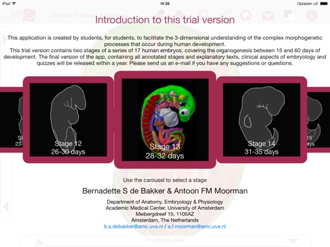
The 3D Atlas of Human Embryology - Lite, by Bernadette S de Bakker, MSc, Jaco Hagoort and Prof. Dr. Antoon FM Moorman, is an embryology resource that provides clear three-dimensional (3D) insight in human embryology, covering the organogenesis between 15 and 60 days of development. This free Lite version contains two human embryonic specimens (stage 8 and 13) from the Carnegie Collection. Every stage is reconstructed in 3D, which enables the user to interactively zoom in and out on the embryo and to show every structure in a transparent fashion in order to enhance the understanding of this difficult topic. Besides the 3D models, every stage contains additional embryonic and clinical information, label quizzes, multiple-choice quizzes and educational views. The full version of the app, containing all 17 annotated stages will be released soon.
This atlas was created by students and embryologists of the Department of Anatomy, Embryology & Physiology of the Academic Medical Center (AMC) in Amsterdam, the Netherlands. The 3D models are manually created based on stacks of histological sections of real human embryos, mainly from the Carnegie Collection. The current inaccessibility of the collection makes this atlas even more valuable.
Uses
This application can be used as a complement to academic learning tools in the (bio)medical curriculum. The bookmark function enables embryology and biology teachers but also scientists to create unlimited instructive 3D figures of the models. This application can also be used by medical professionals and physicians, especially in the field of gynecology, pediatrics and clinical genetics.
Features
• This application is in English.
• Stage selection – Learn embryology based on subsequent human embryonic stages.
• Actual size monitor – Get a grip on the actual size of the embryo you are viewing.
• Structure tree – Every structure is arranged in a structure tree, functionally divided into organ systems.
• Interactive models – Rotational models of real human embryos are provided. Every structure is annotated and can be turned on, off or be shown in a transparent fashion.
• Educational texts – Explanatory texts with clinical aspects were written about every structure. The used terminology is in accordance with the Terminologia Embryologia (2009).
• Search function – When more than two characters are given, the search function gives a list of potential key words which enables the user to efficiently search through the information given in this application.
• Educational views – Typical educational views were selected per stage, for example of the neurenteric canal in the stage 8 embryo and of the developing arterial arches in the stage 13 embryo.
• Bookmark function – Use the bookmark function to save views for later moments, lectures and discussions with fellow scientists, students and patients.
• Label quizzes – Train your knowledge of the topography of all embryonic structures with the label quizzes.
• Multiple-choice quizzes – Train your knowledge of the embryology via targeted multiple choice quizzes. This Lite version contains a multiple-choice quiz of the stage 8 embryo as an example for this feature in the full version.
• Free periodic updates
Contents
Integumentary system - Skeletal and muscular system - Alimentary system - Respiratory system - Urogenital system - Coelom - Endocrine system - Nervous system - Sense organs


